
Multiclass boosting SVM using different texture features in HEp-2 cell
staining pattern classification
Kuan Li Jianping Yin
National University of Defense Technology, Changsha, China
{likuan, jpyin}@nudt.edu.cn
Zhi Lu Xiangfei Kong Rui Zhang Wenyin Liu*
City University of Hong Kong
{luzhi2, xfkong2}@student.cityu.edu.hk, {rzhang22, csliuwy}@cityu.edu.hk
Abstract
In this paper, we present four image descriptors for
HEp-2 cell staining patterns classification, including
LBP, Gabor, DCT, and a global appearance statistical
descriptor. A multiclass boosting SVM algorithm is pro-
posed to integrate these descriptors together: (1) within
each boosting round, four multiclass posterior proba-
bility SVMs are trained corresponding to four descrip-
tors, and then combined to an integrated classifier; (2)
AdaBoost.M1 is modified to enhance the performance
of the integrated classifiers. Experimental results over
721 images with 5-fold cross validation show the pro-
posed method is effective and can improve the classifi-
cation accuracy.
1. Introduction
Anti-nuclear antibodies (ANAs), which are autoan-
tibodies directed against contents of the cell nucleus,
have been detected in the serum of patients with many
autoimmune diseases. The recommended method for
ANA testing is the indirect immunofluorescence (IIF)
based on HEp-2 substrate [8]. Currently, the identi-
fication of IIF slides is manually inspected by physi-
cians with a microscope. However, manpower-based
IIF slides analysis is a tedious, time-consuming and
error-prone job. The vast amount of image data and the
lack of physician work forces make things worse.
There are four main steps in the IIF diagnostic proce-
dure, namely image acquisition, mitosis detection, flu-
orescence intensity classification and staining pattern
recognition. The last step is very challenging and im-
portant since several different patterns may match with
different autoimmune diseases. Therefore, Computer-
Aided Diagnosis (CAD) system would bring significant
benefits to overcome these limitations. Staining patterns
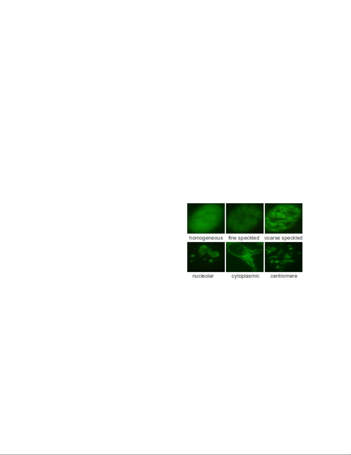
are classified into one of the following six groups: ho-
mogeneous, fine speckled, coarse speckled, nucleolar,
cytoplasmic and centromere. Examples of the above
defined patterns are shown in Fig. 1.
Figure 1. Examples of different staining
patterns.
Early work for HEp-2 cell classification are given in
[1, 6]. The datasets used in [1] and [6] consisted of 1041
and 321 fluorescence cells, respectively. Based on the
decision tree classifier and texture features computed on
segmented cells, their systems exhibited an error rate of
16.9% [1] and 25.6% [6]. More detailed reviews can be
found in [7].
This paper focuses on the classification of pre-
segmented HEp-2 cell staining patterns. We fully ex-
ploit the texture features of HEp-2 cells based on LBP,
Gabor and DCT. Furthermore, a global appearance sta-
tistical feature extraction method is introduced. Finally,
a multiclass boosting SVM algorithm is proposed to in-
corporate different information together to achieve bet-
ter classification performance.
下载后可阅读完整内容,剩余3页未读,立即下载

weixin_38727579
- 粉丝: 4
- 资源: 918
 我的内容管理
展开
我的内容管理
展开


最新资源
- C++标准程序库:权威指南
- Java解惑:奇数判断误区与改进方法
- C++编程必读:20种设计模式详解与实战
- LM3S8962微控制器数据手册
- 51单片机C语言实战教程:从入门到精通
- Spring3.0权威指南:JavaEE6实战
- Win32多线程程序设计详解
- Lucene2.9.1开发全攻略:从环境配置到索引创建
- 内存虚拟硬盘技术:提升电脑速度的秘密武器
- Java操作数据库:保存与显示图片到数据库及页面
- ISO14001:2004环境管理体系要求详解
- ShopExV4.8二次开发详解
- 企业形象与产品推广一站式网站建设技术方案揭秘
- Shopex二次开发:触发器与控制器重定向技术详解
- FPGA开发实战指南:创新设计与进阶技巧
- ShopExV4.8二次开发入门:解决升级问题与功能扩展
资源上传下载、课程学习等过程中有任何疑问或建议,欢迎提出宝贵意见哦~我们会及时处理!
点击此处反馈


